Microscopy and Computed Tomography

  • Manuf.: Zeiss
  • e--source: Schottky field emission gun
  • Accelerating voltage: 0.2–30 kV
  • High resolution imaging with SE, BSE and InLens-detectors down to 3 nm
  • Magnification: 20X–900,000X
  • 5-axis sample stage enables tilting and rotation
  • Element analysis by EDX (Bruker XFlash 4010)
  • Analysis of grain orientations and structures by EBSD (Bruker e-FlashHR+)
  • Investigations of magnetic samples possible
  • Extensions: heating table up to  500 °C, DDS32 tensile/compression module (K&W), TKD head for EBSD detector
  • Manuf.: Keyence
  • Magnification: 20X–2500X
  • Resolution: 2048 x1536 pixels
  • Fast measurement of objects and features
  • Automatic particle measurement
  • Generation of 3-dim. areas or 3D models incl. altitude profile
  • Software-side image enhancement through gloss removal, HDR, etc.
  • Illumination and viewing from different angles by tilting camera head

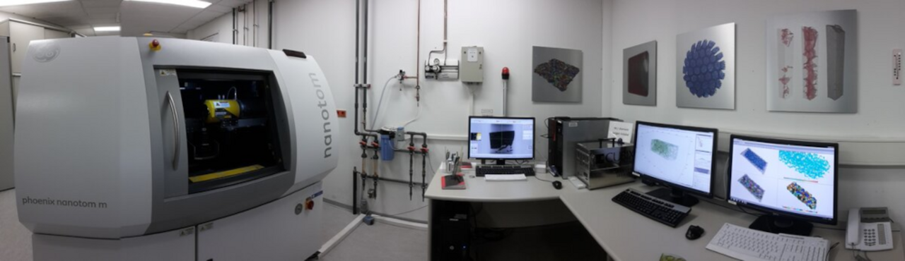
X-ray absorbtion CT is a non-destructive imaging method to analyze the inner constitution of solid objects and allows to distinguish between different phases (including defects and pores) due to their different absorbtion properties.

  • 180kV Tungsten X-ray tube with macro- and nanofocus mode (for smaller focal spot sizes and higher resolutions)
  • Al- and Cu-Filter (0.2 to 1 mm)
  • 2D amorphous silicon flat panel detector (DXR500L), 3070x2400 pixel (100µm pixel pitch), CsI scintillator, 500 ms minimum exposer time
  • Magnification (geometric-optical) up to 300x
  • Voxelsize up to 500 nm (commonly 2 – 6 µm)
  • Sample size (diameter): up to 5 mm Fe-based, up to 20 mm Al-based
  • Resolution max. 0.5 µm
  • Scanmodi: Sectionscan, Fastscan, Singlescan, Multiscan
  • Software: GE datos Acquisition 2.0, GE datos Reconstruction 2.0 (with beam hardening correction algorithm), Visual Studio VG Studio max 2.2, FEI AvizoFire 8.1 & 9